The procedure for in-gel digestion of protein spots from Coomassie Blue stained gels was carried out as described in [ ]. Dataset of protein species from human liver Article Snippet: Proteomic and phosphoproteomic analysis of polyethylene glycol-induced osmotic stress in root tips of common bean Phaseolus vulgaris L. Proteomic analysis and candidate allergenic proteins in Populus deltoides CL. The relative volume of each spot was determined from the spot intensities in pixel units and normalized to the sum of the intensities of all the spots of the gel. The relative abundance of each protein was then calculated by normalizing the volume of a protein target to the sum of volume of all protein spots on the same 2-D gel image. For protein carbonylation profiles, the electrophoresed gels were transferred to PVDF membrane and carbonylated protein spots were detected as mentioned above.
| Uploader: | Vojas |
| Date Added: | 14 October 2013 |
| File Size: | 47.86 Mb |
| Operating Systems: | Windows NT/2000/XP/2003/2003/7/8/10 MacOS 10/X |
| Downloads: | 93134 |
| Price: | Free* [*Free Regsitration Required] |
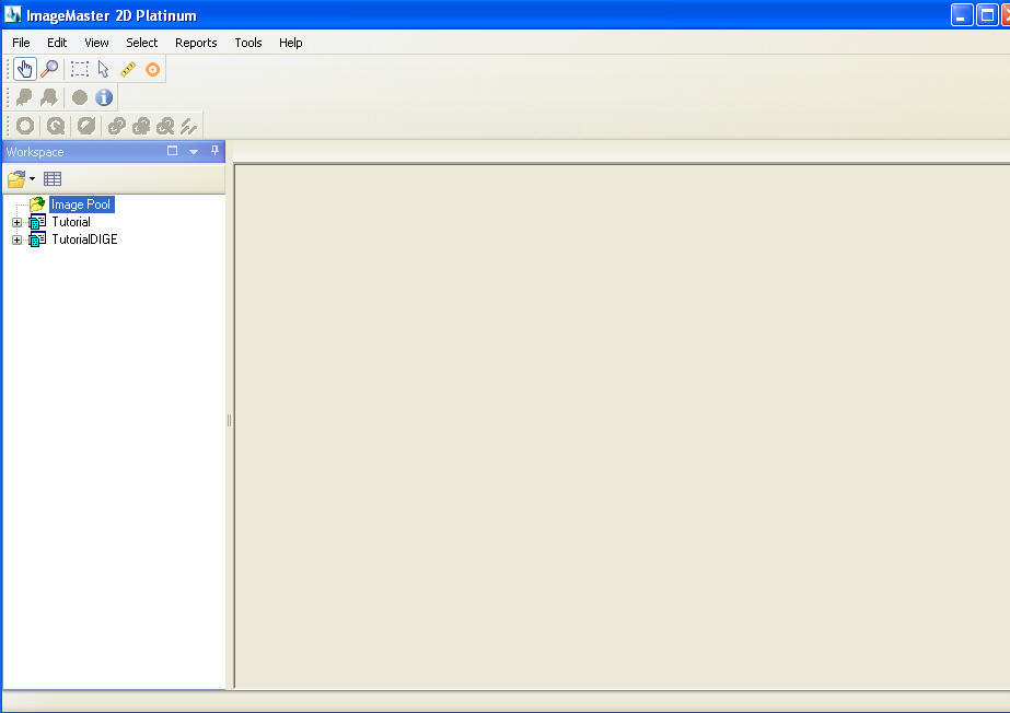
ImageMaster 2D Platinum Download -
The gel produced was stained with Coomassie Brilliant Blue R The linear ramping mode of the IEF voltage was used as follows; 30 V for 6 h, 60 V for 6 h, V for 1 h, V for 1 h, and V for 1 h, followed by V for approximately 7. Gels three of each imagemasterr then silver-stained and scanned, and the resulting images were analyzed using the ImageMaster 2D Platinum software for spot detection, quantification, and comparative and statistical analyses as described in the Kmagemaster section.
Western blots were used to validate the imqgemaster 2D proteomic analysis for the antioxidant enzymes. Proteomic analysis 66.0 candidate allergenic proteins in Populus deltoides CL. Following zymography, the degree of gelatin digestion was quantified using ImageMaster 2D Platinum softwareVersion 5. Quantification of Enzymatic Activity Journal of Experimental Botany Article Title: The green labels identify proteins whose expression is altered by a factor of greater than 1.
Expressing, Electrophoresis, Incubation, Staining, Software. Two-dimensional Polyacrylamide Gel Electrophoresis A proteomic analysis of mushroom polysaccharide-treated HepG2 cells Article Snippet: Hsp90 inhibitor allylaminodemethoxygeldanamycin inhibits the proliferation of ARPE cells.
Thereafter, the resolved proteins were stained with Deep Purple protein fluorescence dye GE Healthcare and visualized by using Typhoon laser scanner GE Healthcare. Following electrophoresis, all gels were stained with colloidal Coomassie brilliant blue and scanned with a calibrated densitometer ImageScanner, GE Healthcare. Staining was performed by Coomassie Brilliant Blue R Following electrophoresisall kmagemaster were stained with colloidal Coomassie brilliant blue and scanned with a calibrated densitometer ImageScanner, GE Healthcare.
Silver-stained gels were scanned using an Atrix scan plus Microtek, Taiwan, Imagemxsterand the resulting images were analyzed using the ImageMaster 2D Platinum software Amersham Bioscience for spot detection, quantification, and comparative and statistical analyses.
Journal of Biomedical Science doi: Attached and floating cells were harvested as described in the immunoblot analysis, and cell pellets were lysed in 2D lysis buffer at room temperature for 1 h. The procedure for in-gel digestion imagemastee protein spots from Coomassie Blue stained gels was carried out as described in [ ].

Gel portion showing differential expression of spot ID84 and ID85 of hypobaric hypoxia exposed Sprague Dawley rats; a tolerant: Sample preparation and 2-DE Hepatic protein Carbonylation profiles induced by lipid accumulation and oxidative stress for investigating cellular response to non-alcoholic fatty liver disease in vitro Article Snippet: The relative volume of circle-indicated spot was analyzed by ImageMaster 2D Platinum software.
The mean and SD normalized volume of each protein spot were calculated, and statistical comparisons between the intensity of the control and the AAG treated spots were conducted using Student't-test with p Article Title: The apparent molecular weight of each protein in the gel was determined using protein markers. Dataset of protein species from human liver Journal: The location of gelatinolytic activity was detectable as clear bands against the blue background of stained gelatin.
The image was digitally inverted, so that the integration of bands was reported as positive values.

Strips were then equilibrated at room temperature for 15 imagemadter in 10 ml equilibration solution [6 M urea, 50 mM Tris-HCl pH8. The relative abundance of each protein was then calculated by normalizing the volume of a protein target to the sum of volume of all protein spots on the same 2-D gel image.
Imagemaster 2d platinum 6.0
Two replicate gels were run for each of three different pooled pollen samples collected from different batches of plants. Accession numbers and fold changes of the identified proteins from proteomic analysis were input as a dataset in Ingenuity Pathway Analysis software Qiagen, Germantown, MD, USA to predict the relevant molecular networks.
Treatment-affected spots were marked by arrows and numbered consecutively. The relative volume of each spot was determined from the spot intensities in pixel units and normalized to the sum of the intensities of all the spots of the gel.
The resulting peak list files were submitted to http:

Комментариев нет:
Отправить комментарий